


Meniscus tear (knee cartilage damage)
A meniscus tears in two quite different ways. In a young person it takes a sharp twist on a loaded leg — football, an awkward landing, a change of direction.

Prescription request online
A doctor will review your case and issue a prescription if medically appropriate.

Talk to a doctor online
Discuss your symptoms and possible next steps with a doctor online.
On this page
Medicines commonly prescribed for Meniscus tear (knee cartilage damage)
For informational purposes only. Always consult a doctor before using any medicine.
 IBUPROFEN TIVA 400 mg SOFT GEL CAPSULESIllustrative imageDosage form: CAPSULE, 400 mgActive substance: ibuprofenManufacturer: Teva Pharma S.L.U.Prescription not required
 PARACETAMOL MABO-FARMA 1 g TABLETSIllustrative imageDosage form: TABLET, 1 gActive substance: paracetamolManufacturer: Mabo Farma S.A.Prescription required
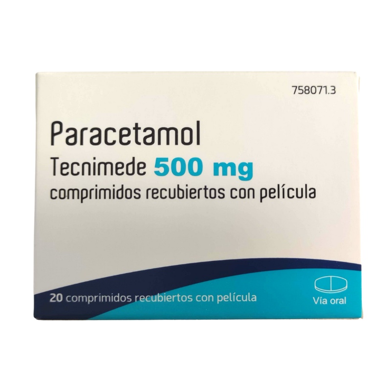 PARACETAMOL PHARMACLAN 500 mg FILM-COATED TABLETSIllustrative imageDosage form: TABLET, 500 mgActive substance: paracetamolManufacturer: Pharmaclan S.R.O.Prescription not required
A meniscus tears in two quite different ways. In a young person it takes a sharp twist on a loaded leg — football, an awkward landing, a change of direction. From the forties onwards it usually happens the other way round: the knee starts clicking and aching with no injury anyone can recall. Which of the two you are dealing with decides almost everything that follows, from the tests you need to whether surgery makes any sense at all.
What the meniscus is and why it tears
Between the thigh bone and the shin bone sit two crescent-shaped pads of cartilage, the inner and the outer meniscus. They spread body weight across the joint, deepen the flat top of the shin bone under the rounded end of the thigh bone, and steady the knee when it rotates. Without them the load would gather at a single point.
A fresh tear happens when the foot stays planted and the body turns: with the knee bent, the meniscus is pinned between the bones and cannot slip out of the way in time. That is how menisci tear in sport and in falls.
The second story belongs to age. Over the years the cartilage loses water and turns brittle, so a split appears during an everyday movement — standing up from a deep squat, climbing out of a car, turning with a bag in hand. Often the person cannot tie the pain to any moment at all. The same changes turn up in people with no complaints whatsoever, so a split seen on a scan does not by itself explain the pain.
The meniscus heals poorly, and not everywhere. Blood vessels reach only the outer rim, near the joint capsule; the inner part lives on joint fluid and has nothing to knit with. A tear at the edge therefore has a chance of healing on its own or after stitching, while a tear deep inside does not.
What the knee feels like
The pain is usually pinpoint and sits along the joint line, on the inner or outer side, exactly at the level of the gap between the bones. You can find it with one finger.
- swelling that builds up not at once but over several hours or by the next day;
- clicking, grating, a sense of something rolling or catching inside;
- the knee gives way for an instant when you step or use stairs;
- difficulty straightening the leg fully, squatting or kneeling;
- going down stairs is worse than going up.
With a wear-related split the picture is quieter: a dull ache and mild puffiness that show up after a long walk or a stretch of work on your haunches and settle within a couple of days.
Locking deserves separate attention: a detached flap jams between the bones, the knee sticks half bent and will not straighten at all. This is not merely severe pain, it is a mechanical block, and it calls for an orthopaedic assessment rather than a wait-and-see approach.
What to do in the first few days
For the first two or three days you cut the load, but you do not switch the leg off: an immobile knee loses muscle quickly and recovers worse afterwards. Walk as much as you can without the pain rising, using a stick if that helps.
- cold through a cloth for 15 to 20 minutes several times a day, never ice straight against the skin;
- an elastic bandage or a knee support during the day, removed for the night;
- the leg on a cushion above the level of the heart when you are lying down;
- paracetamol for pain; a short course of anti-inflammatory painkillers also fits, provided you have no ulcer, no kidney disease and no advice against them from your doctor.
In those first days avoid heating the knee, hot baths and saunas, warming rubs and kneading the joint with your hands — heat and massage make the swelling worse. Do not try to force a locked knee straight. Return to activity gradually and let the swelling be your guide: if the knee fills up again after a session, you have taken on too much.
When to see a doctor and when to go to emergency care
Straight away, calling an ambulance on the single European number 112 or getting to emergency care without driving yourself:
- the knee is deformed, or the kneecap or the joint is clearly out of place;
- there was a crack at the moment of injury and you cannot put any weight on the leg;
- the joint filled up tight within the first hour after the injury, which usually means blood and therefore a torn ligament or a fracture;
- the foot has gone numb, pale or cold, or you cannot feel your toes;
- a wound that runs into the joint;
- the knee is hot, red and sharply painful with chills or fever. Infection inside a joint destroys cartilage within days, and here the count is in hours.
Within the next few days, rather than in a month's time, you need an orthopaedic assessment if the knee has locked and will not straighten, if it repeatedly gives way when you walk, or if the swelling returns after every effort.
A planned appointment is warranted when the pain lasts more than two or three weeks despite rest, keeps you awake or interferes with ordinary tasks, and also if the knee stays stiff for more than half an hour in the morning.
How the diagnosis is made
It all starts with a conversation about the mechanism: what the leg was doing when the pain struck, whether there was a sense of a click, how fast the swelling appeared. The doctor then presses along the joint line and performs rotation and bending manoeuvres — with a damaged meniscus these reproduce the familiar pain or a catch. Such tests point the thinking in a direction, but on their own they prove nothing.
An X-ray does not show the meniscus, because cartilage simply is not visible on it. The film is taken for another reason: to rule out a fracture and to see how far the joint space has narrowed, that is, whether arthritis explains the pain better than a split does.
The tear is confirmed by magnetic resonance imaging. It shows the meniscus itself, the shape and position of the tear, and along with it the ligaments and the bone, which are often injured at the same time. Ultrasound is not up to this job: it picks up fluid and cysts but misses tears in the depth of the tissue.
Arthroscopy, looking inside the joint with a camera, is diagnosis and treatment at once, but it is not used as a first step. And one important caveat: from the forties onwards, meniscal changes are found on scans in a large share of people who have no symptoms. The person and the complaints are treated, not the picture.
How it is treated
Most people need no operation. A fresh tear without locking, and almost any age-related split, are treated with loading: a programme of exercises for the quadriceps, the glutes and balance, guided by a physiotherapist. It runs for six to twelve weeks, and it is not a matter of waiting for the pain to pass but of steady work with a gradual rise in demand.
Wear-related splits in particular have been studied with unusual care: removing part of the meniscus by arthroscopy in middle-aged and older people gave no advantage over a properly supervised exercise programme. If the knee does not lock, there is no hurry towards the operating theatre.
Surgery is needed when there is a mechanical problem: a locked knee, a large tear whose flap shifts into the joint, a leg that repeatedly gives way, or honest rehabilitation that has failed. There are two options. Stitching the meniscus preserves tissue and is preferred when the tear is recent and lies in the zone with a blood supply, though it demands a long, careful regime afterwards. Removing the damaged part relieves symptoms sooner but leaves less tissue behind.
Neither tablets nor injections make torn cartilage knit together. Cartilage supplements have no effect on a tear, and a steroid injection may settle inflammation but does not solve a mechanical block.
Recovery and what comes later
Treated without surgery, a knee usually settles over a period of a few weeks to three months. After partial removal people walk almost immediately and return to sport at around six weeks. After a repair the regime is stricter: limits on bending and weight-bearing, crutches, a brace, and full activity only after several months. That gap in timing is the price paid for the tissue that was saved.
In the long run what matters most is how much meniscus remains: the less was taken out, the lower the risk of knee arthritis over the years. That risk is also shaped by things within your control, namely body weight and the strength of the thigh muscles.
After an operation, contact a doctor without delay if you develop a fever, if the joint becomes increasingly swollen and hot, if the calf is painful and swollen, or if sudden breathlessness or chest pain appears.
Online consultation
A remote appointment answers the first and central question well: what you are dealing with and where to go. From the mechanism of the injury, the speed at which the swelling appeared and the presence of locking or clicking, a doctor can judge whether this looks like a meniscus, whether imaging is needed and in what order. Record a short video of yourself bending and straightening the knee, and measure the circumference of both legs at the same point: the difference in centimetres speaks both of swelling and of lost muscle.
The conversation is useful later on too: making sense of a scan report, weighing up an operation that has been offered, telling apart what a repair achieves and what removal achieves, and setting out an exercise plan for the coming weeks. The limits are obvious: a joint cannot be palpated or tested through a screen, and a surgical decision is taken after a hands-on examination.
This material is for information only and does not replace medical advice.
